Tuesday, 29 April 2014

How is Lung Cancer Diagnosed and Treated?

Testing healthy people for lung cancer
People with an increased risk of lung cancer consider annual computerized tomography (CT) scans to look for lung cancer. If you're 55 or older and smoke or used to smoke, talk with your doctor about the benefits and risks of lung cancer screening.
Some studies show lung cancer screening saves lives by finding cancer earlier, when it may be treated more successfully. But other studies find that lung cancer screening often reveals more benign conditions that may require invasive testing and expose people to unnecessary risks and worry.
Tests to diagnose lung cancer
If there's reason to think that you may have lung cancer, your doctor can order a number of tests to look for cancerous cells and to rule out other conditions. In order to diagnose lung cancer, your doctor may recommend:
·         Imaging tests. An X-ray image of your lungs may reveal an abnormal mass or nodule. A CT scan can reveal small lesions in your lungs that might not be detected on an X-ray.
·         Sputum cytology. If you have a cough and are producing sputum, looking at the sputum under the microscope can sometimes reveal the presence of lung cancer cells.
·         Tissue sample (biopsy). A sample of abnormal cells may be removed in a procedure called a biopsy.
Your doctor can perform a biopsy in a number of ways, including bronchoscopy, in which your doctor examines abnormal areas of your lungs using a lighted tube that's passed down your throat and into your lungs; mediastinoscopy, in which an incision is made at the base of your neck and surgical tools are inserted behind your breastbone to take tissue samples from lymph nodes; and needle biopsy, in which your doctor uses X-ray or CT images to guide a needle through your chest wall and into the lung tissue to collect suspicious cells.
A biopsy sample may also be taken from lymph nodes or other areas where cancer has spread, such as your liver.

Lung cancer staging
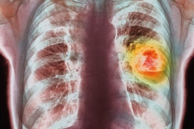
Once your lung cancer has been diagnosed, your doctor will work to determine the extent (stage) of your cancer. Your cancer's stage helps you and your doctor decide what treatment is most appropriate.
Staging tests may include imaging procedures that allow your doctor to look for evidence that cancer has spread beyond your lungs. These tests include CT scans, magnetic resonance imaging (MRI), positron emission tomography (PET) and bone scans. Not every test is appropriate for every person, so talk with your doctor about which procedures are right for you.
Stages of lung cancer
·         Stage I. Cancer is limited to the lung and hasn't spread to the lymph nodes. The tumor is generally smaller than 2 inches (5 centimeters) across.
·         Stage II. The tumor at this stage may have grown larger than 2 inches, or it may be a smaller tumor that involves nearby structures, such as the chest wall, the diaphragm or the lining around the lungs (pleura). Cancer may also have spread to the nearby lymph nodes.
·         Stage III. The tumor at this stage may have grown very large and invaded other organs near the lungs. Or this stage may indicate a smaller tumor accompanied by cancer cells in lymph nodes farther away from the lungs.
·         Stage IV. Cancer has spread beyond the affected lung to the other lung or to distant areas of the body.
Small cell lung cancer is sometimes described as being limited or extensive. Limited indicates cancer is limited to one lung. Extensive indicates cancer has spread beyond the one lung.


The treatments for lung cancer depend on the stage of the cancer, personal characteristics, health status, type of the cancer and age. A number of therapies are provided to a patient as there is no single treatment available for lung cancer. Radiation, surgery and chemotherapy are considered as the major lung cancer treatment.
  • Surgery : One of the oldest methods for treating lung cancer is surgery. The surgical removal of the tumor and surrounding lymph nodes is done if there is I or II stage cancer that has not metastasized. Palliative or curative are the two types of lung cancer surgeries. Palliative surgery may not remove cancer but can remove an open airway or obstruction that was making the patient uncomfortable. Curative surgery removes all types of cancerous tissue in those patients who are in early stage lung cancer.
  • Chemoembolization:Strong chemicals are used in chemotherapy that interferes with cell division process and damages DNA or proteins. The aim of these treatments is to rapidly divide the cells. The normal cells can be recovered from any chemical-induced damage whereas cancer cells cannot be recovered. The medicines in chemotherapy travel in a systematic way by passing from the complete body and destroying the original tumor cells that have spread in the whole body. Usually many therapies are combined that also includes many types of chemotherapy
  • Radiation Therapy :Radiation therapy may be employed as a treatment for both NSCLC and SCLC. Radiation therapy uses high-energy X-rays or other types of radiation to kill dividing cancer cells. Radiation therapy may be given as curative therapy, palliative therapy (using lower doses of radiation than with curative therapy), or as adjuvant therapy in combination with surgery or chemotherapy. Radiation therapy generally only shrinks a tumor or limits its growth when given as a sole therapy, yet in 10%-15% of people it leads to long-term remission and palliation of the cancer. Combining radiation therapy with chemotherapy can further prolong survival when chemotherapy is administered. External radiation therapy can generally be carried out on an outpatient basis, while internal radiation therapy requires a brief hospitalization.
  • Targeted Therapy :Targeted therapy drugs more specifically target cancer cells, resulting in less damage to normal cells than general chemotherapeutic agents. Erlotinib and gefitinib target a protein called the epidermal growth factor receptor (EGFR) that is important in promoting the division of cells. This protein is found at abnormally high levels on the surface of some types of cancer cells, including many cases of non-small cell lung cancer.

 For more information visit:          http://www.medworldindia.com         
                    https://www.facebook.com/medworld.india

Please scan and email your medical reports  to us at care@medworldindia.com and we shall get you a Free Medical Opinion from India’s Best Doctors.

Call Us : +91-9811058159
Mail Us : care@medworldindia.com

Sunday, 27 April 2014

Frequently Asked Questions About Stomach Cancer ?


A. The stomach is a sack-like organ located just under the diaphragm (muscle under the lungs). Stomach cancer, also called gastric cancer, is the name for cancer that begins in the stomach, generally the stomach lining. This type of cancer can eventually spread to lymph nodes and organs such as the liver, pancreas, colon, lungs and ovaries. People occasionally confuse the stomach organ with the abdominal area, saying they have a "stomach ache" when really the pain could be occurring in the appendix, small intestine, colon (large intestine) or gall bladder, along with the actual stomach. The stomach can be divided into five sections, and the location of the cancer in the stomach can affect things like symptoms, prognosis and treatment options.

Q. What are the Causes and Risk Factors for Stomach Cancer?

A. Risk factors are conditions that increase a person’s chance of getting a type of cancer. Risk factors are different for various types of cancer. Doctors have determined a number of risk factors for stomach cancer:
  • Aging – There is a sharp increase in stomach cancer after 50. Most people diagnosed with stomach cancer are in the 60-70 age range.
  • Being male – Stomach cancer is about two times more common in men than in women.
  • Diet – Foods that are smoked, salted fish and meat, pickled vegetables, and foods that are at the same time high in starch and low in fiber have been identified as possible risk factors. Scientists believe that the dramatic decline in stomach cancer incidence in the United States from the 1930s may be due to the increased use of refrigeration for food storage, which replaced salting or smoking food for storage.
  • Tobacco and alcohol abuse – Increases the risk of cancers in the upper portion of the stomach, which can be difficult to treat successfully.
  • Previous stomach surgery – After surgery, more nitrite-producing bacteria are present in the stomach. Nitrites can be converted by other bacteria into compounds that have been found to cause stomach cancer in animals.
  • Family history of stomach cancer – Several close blood relatives who have or had stomach cancer increases a person’s risk.
  • Helicobacter pylori infection – An infection that, if long-term, can lead to chronic atrophic gastritis, which is inflammation of the stomach’s inner layer. Chronic atrophic gastritis is a possible precancerous change to the lining of the stomach.
  • Also, risk may be increased, to varying degrees, for people with pernicious anemia, achlorhydria, Menetrier’s disease, familial cancer syndromes, stomach polyps and blood group A.


A. These symptoms might be caused by stomach cancer:
  • Indigestion or a burning sensation (heartburn)
  • Discomfort or pain in the abdomen
  • Nausea and vomiting
  • Diarrhea or constipation
  • Bloating of the stomach after meals
  • Loss of appetite
  • Weakness and fatigue
  • Bleeding (vomiting blood or having blood in the stool)
However, many of these symptoms are more often caused by other medical conditions. It is important to consult a physician to find out what is causing symptoms.


A . Surgery : The goal of surgery is to remove all of the stomach cancer and a margin of healthy tissue, when possible. Options include:


    • Removing a Portion of the Stomach (Subtotal Gastrectomy).During subtotal Gastrectomy, the surgeon removes only the portion of the stomach affected by cancer. Subtotal gastrectomy can be of two types.


    • Distal subtotal gastrectomy is performed if the tumor cells are present in the lower part of the stomach near to the stomach duodenum junction.
    • Proximal subtotal gastrectomy is done when the tumor is situated in the upper part of the stomach and also involves the esophagus


    • Removing the Entire Stomach (Total Gastrectomy).Total Gastrectomy involves removing the entire stomach and some surrounding tissue. The esophagus is then connected directly to the small intestine to allow food to move through your digestive system.


    • Removing Lymph nodes to look for Cancer. The surgeon examines and removes lymph nodes in your abdomen to look for cancer cells.

  • Chemotherapy : This therapy uses cancer destroying drugs for treating stomach cancer. When the cancer has invaded the stomach wall layers surrounding the lymph nodes and nearby organs then chemotherapy is given. For shrinking the tumor first, chemotherapy is given before the surgery. It can also be given after the surgery for destroying the remaining cancer cells. Chemotherapy can also be combined with radiation therapy that provides relieves from many cancer symptoms and can also delay the recurrence of the cancer.
  • Radiation therapy : Doctors may recommend radiation therapy in combination with chemotherapy or surgery. Radiation oncologists a have extensive experience in the treatment of gastric cancer, and they use specialized techniques to limit the radiation dose to surrounding healthy tissue.

 For more information visit:          http://www.medworldindia.com         
                    https://www.facebook.com/medworld.india

Please scan and email your medical reports  to us at care@medworldindia.com and we shall get you a Free Medical Opinion from India’s Best Doctors.

Call Us : +91-9811058159
Mail Us : care@medworldindia.com

Friday, 25 April 2014

Prostate Cancer What all Men Must Know - Symptons , Diagnosis , Stages and Treatment in India

The prostate is part of a man's reproductive system and is a walnut-sized gland located between the bladder and the penis. The prostate is just in front of the rectum. The urethra runs through the center of the prostate, from the bladder to the penis, letting urine flow out of the body. The prostate is a gland.
The  prostate cancer is the most common type of cancer in men. Prostate cancer is the second leading cause of cancer death in men, exceeded only by lung cancer. Adenocarcinoma of the prostate is the clinical term for a cancerous tumor of the prostate gland. As prostate cancer grows, it may spread to the interior of the gland, to tissues near the prostate, to sac-like structures attached to the prostate (seminal vesicles), and to distant parts of the body . Prostate cancer confined to the gland often is treated successfully.

Symptoms of Prostate Cancer

A man with prostate cancer may not have any symptoms. Symptoms of prostate cancer are often similar to those of benign prostatic hyperplasia (BPH). Men observing the following signs and/or symptoms should see their physician for a thorough examination:
·         Urinary problems -  Not being able to pass urine  Having a hard time starting or stopping the urine flow
·         Needing to urinate often, especially at night
·         Weak flow of urine
·         Urine flow that starts and stops
·         Pain or burning during urination
·         Blood in the urine or semen

·         Frequent pain in the lower back, hips, or upper thighs If you have any of these symptoms, you should tell your doctor so that problems can be diagnosed and treated.


Blood test for prostate-specific antigen (PSA): A lab checks the level of PSA in your blood sample.
The prostate makes PSA. A high PSA level is commonly caused by BPH or prostatitis (inflammation of the prostate). Prostate cancer may also cause a high PSA level. The digital rectal exam and PSA test can detect a problem in the prostate. However, they can't show whether the problem is cancer or a less serious condition. If you have abnormal test results, your doctor may suggest other tests to make a diagnosis.

Transrectal ultrasound: The doctor inserts a probe into the rectum to check your prostate for abnormal areas. It also measures the size of the prostate, which can help to determine if the PSA level is elevated for the size of the prostate. The probe sends out sound waves that people cannot hear (ultrasound). The waves bounce off the prostate. A computer uses the echoes to create a picture called a sonogram.

Transrectal biopsy: A biopsy is the removal of tissue to look for cancer cells. It's the only sure way to diagnose prostate cancer. The doctor inserts needles through the rectum into the prostate. The doctor removes small tissue samples (called cores) from many areas of the prostate. Transrectal ultrasound is usually used to guide the insertion of the needles. A pathologist checks the tissue samples for cancer cells.

If cancer cells are found, the pathologist studies tissue samples from the prostate under a microscope to report the grade of the tumor. The grade tells how much the tumor tissue differs from normal prostate tissue. It suggests how fast the tumor is likely to grow. Tumors with higher grades tend to grow faster than those with lower grades. They are also more likely to spread. Doctors use tumor grade along with your age and other factors to suggest treatment options. The most commonly used system for grading is the Gleason score. Gleason scores range from 2 to 10. To come up with the Gleason score, the pathologist uses a microscope to look at the patterns of cells in the prostate tissue

Staging of Prostate Cancer

If the biopsy shows that you have cancer, your doctor needs to learn the extent (stage) of the disease to help you choose the best treatment. Staging is a careful attempt to find out whether the tumor has invaded nearby tissues, whether the cancer has spread and, if so, to what parts of the body. Some men may need tests that make pictures of the body:

 Bone scan: The doctor injects a small amount of a radioactive substance into a blood vessel. It travels through the bloodstream and collects in the bones. A machine called a scanner detects and measures the radiation. The scanner makes pictures of the bones on a computer screen or on film. The pictures may show cancer that has spread to the bones. Many times a plain x-ray is taken to help evaluate an abnormality seen on a bone scan.

 CT scan: An x-ray machine linked to a computer takes a series of detailed pictures of your pelvis or other parts of the body. Doctors use CT scans to look for prostate cancer that has spread to lymph nodes and other areas. You may receive contrast material by injection into a blood vessel in your arm or hand, or by enema. The contrast material makes abnormal areas easier to see.

 MRI: A strong magnet linked to a computer is used to make detailed pictures of areas inside your body. The doctor can view these pictures on a monitor and can print them on film. An MRI can show whether cancer has spread to lymph nodes or other areas.
When prostate cancer spreads, it's often found in nearby lymph nodes. If cancer has reached these nodes, it also may have spread to other lymph nodes, the bones, or other organs. When cancer spreads from its original place to another part of the body, the new tumor has the same kind of abnormal cells and the same name as the primary tumor. For example, if prostate cancer spreads to bones, the cancer cells in the bones are actually prostate cancer cells.

 Stage I  The cancer can't be felt during a digital rectal exam, and it can't be seen on an imaging study, such as ultrasound. It's found by chance when surgery is done for another reason, usually for BPH. The cancer is only in the prostate and is very low grade (low Gleason score)
 Stage II: o The tumor is more advanced or a higher grade than Stage I, but the tumor doesn't extend beyond the prostate. It may be felt during a digital rectal exam, or it may be seen on a sonogram. It is detected either after a needle biopsy or surgery done for other reasons, i.e. resection of the prostate for benign enlargement.

 Stage III: o The tumor extends beyond the capsule (outer covering) of the prostate. The tumor may have invaded the seminal vesicles, but cancer cells haven't spread to the lymph nodes, bones or other organs.
 Stage IV: o The tumor may have invaded the bladder, rectum, or nearby structures (beyond the seminal vesicles). It may have spread to the lymph nodes, bones, or to other parts of the body.

Treatment of Prostate Cancer in India
Men with prostate cancer have many treatment options. The treatment that's best for one man may not be best for another. Your doctor will make recommendations that are best for each individual. The options include active surveillance (also called watchful waiting), surgery, radiation therapy, cryotherapy, hormone therapy, and chemotherapy. You may have a combination of treatments. The treatment that's right for you depends mainly on your age, the grade of the tumor (the Gleason score), the number of biopsy tissue samples that contain cancer cells, the stage of the cancer, your symptoms, and your general health. Your doctor can describe your treatment choices, the expected results of each, and the possible side effects. You and your doctor can work together to develop a treatment plan that meets your medical and personal needs.

Surgery
Surgery is an option for men with early (Stage I or II) prostate cancer. It's sometimes an option for men with Stage III or IV prostate cancer. Before the surgeon removes the prostate, the lymph nodes in the pelvis may be removed. If prostate cancer cells are found in the lymph nodes, the disease may have spread to other parts of the body. If cancer has spread to the lymph nodes, the surgeon does not always remove the prostate and may suggest other types of treatment. After removing the prostate, the bladder is reconnected to the urethra (tube that men urinate through). Once healed, this will allow men to urinate normally. There are several types of surgery for prostate cancer. Each type has benefits and risks. You and your doctor can talk about the types of surgery and which may be right for you:

 Robotic laparoscopic surgery: The surgeon removes the entire prostate through small cuts  A laparoscope and a robot are used to help remove the prostate. Instruments are passed through the small cuts and are used to remove the prostate. The surgeon uses handles below a computer display to control the robot's arms.

 Open surgery: The surgeon makes a large incision (cut) into your body to remove the tumor. There are two approaches:

Through the abdomen: The surgeon removes the entire prostate through a cut in the abdomen. The incision typically is from the umbilicus (belly button) down to the pelvic bone. This is called a radical retropubic prostatectomy.

Between the scrotum and anus: The surgeon removes the entire prostate through a cut between the scrotum and the anus. This is called a radical perineal prostatectomy.

 Laparoscopic prostatectomy: The surgeon removes the entire prostate through small cuts , rather than a single long cut in the abdomen. A thin, lighted tube (a laparoscope) helps the surgeon see. Other instruments are passed through the small cuts. These instruments are used to remove the prostate.

 TURP: A man with advanced prostate cancer may choose TURP (transurethral resection of the prostate) to relieve symptoms. The surgeon inserts a long, thin scope through the urethra. A cutting tool at the end of the scope removes tissue from the inside of the prostate. TURP may not remove all of the cancer, but it can remove tissue that blocks the flow of urine.
After surgery, a catheter (flexible tube) is often left through the penis into the bladder. This allows the area to heal where the bladder and urethra are reconnected. Oftentimes patients only have to stay in the hospital overnight and can go home the following day. With newer surgical techniques, the complications from surgery are significantly reduced.

Radiation Therapy
Radiation therapy is an option for men with any stage of prostate cancer. Men with early stage prostate cancer may choose radiation therapy instead of surgery. It also may be used after surgery to destroy any cancer cells that remain in the area. It also may be used if the cancer recurs after surgery. In later stages of prostate cancer, radiation treatment may be used to help relieve pain. Radiation therapy (also called radiotherapy) uses high-energy rays to kill cancer cells. It affects cells only in the treated area. Doctors use two types of radiation therapy to treat prostate cancer. Some men receive both types:
  •  External radiation
  •  Internal radiation (implant radiation or brachytherapy)

Hormone Therapy
A man with prostate cancer may have hormone therapy before, during, or after radiation therapy. Hormone therapy is also used alone for prostate cancer that has returned after treatment. Male hormones (androgens) can cause prostate cancer to grow. Hormone therapy keeps prostate cancer cells from getting the male hormones they need to grow. The testicles are the body's main source of the male hormone testosterone. 

Chemotherapy

Chemotherapy may be used for prostate cancer that has spread and no longer responds to hormone therapy. Chemotherapy uses drugs to kill cancer cells. The drugs for prostate cancer are usually given through a vein (intravenous). You may receive chemotherapy in a clinic, at the doctor's office, or at home. Some men need to stay in the hospital during treatment. The side effects depend mainly on which drugs are given and how much.  Most go away when treatment ends.

For more information visit:          http://www.medworldindia.com       
                    
https://www.facebook.com/medworld.india

Please scan and email your medical reports  to us at care@medworldindia.com and we shall get you a Free Medical Opinion from India’s Best Doctors.

Call Us : +91-9811058159
Mail Us : care@medworldindia.com


Wednesday, 23 April 2014

Diagnosis and Treatment for Ovarian Cancer in India

Ovarian Cancer

Ovarian cancer is often termed a "Silent Killer" because its symptoms do not cause alarm amongst the women till it has already reached an advanced stage. Early detection of ovarian cancer offers a 90% cure rate. Sadly, a lack of symptoms from this silent disease means that about 75% of ovarian cancer cases will have spread to the abdomen by the time they are detected.

Symptoms of Ovarian Cancer

The most common symptom of non-Hodgkin's lymphomas is a painless swelling in the lymph nodes of the neck, underarm, or groin. Other symptoms may include fevers, night sweats, tiredness, weight loss, itching, and reddened patches on the skin. Sometimes there is nausea, vomiting, or abdominal pain.


  • Blood tests, including complete blood count (CBC), liver and kidney function test
  • A pelvic exam is performed by the doctors checking the vagina, uterus, ovaries, bladder, and rectum for Endometrial Cancer (Uterine Cancer). An instrument called a speculum is used to widen the vagina so the doctor can see the upper portion of the vagina and the cervix.
  • The Pap Smear test is often performed during a pelvic exam. The doctor uses a wooden scraper (spatula) or small brush to collect a sample of cells from the cervix and upper vagina. The cells are then sent to a medical laboratory to be checked for abnormal changes.
  • A Biopsy may be needed for a definitive diagnosis where the the surgeon performs a laparotomy and removes a sample of tissue. A pathologist examines the tissue to check for cancer cells, hyperplasia, or other conditions.
  • CA-125 assay (for Ovarian Cancer) is a blood test used to measure the level of CA-125, a tumor marker that is often found in higher-than-normal amounts in the blood of women with ovarian cancer.


The main treatments for ovarian cancer are surgery, chemotherapy, and radiation therapy. In some cases two or even all of these treatments will be recommended. 

  • Surgery : Three types of surgeries are involved in treating ovarian cancer such as :–
    • Laparoscopy :Gynaeoncologist perform laparotomy through a large abdominal incision, most often removing your ovaries, uterus, fallopian tubes, nearby lymph glands, a fold of fatty tissue called the omentum and as much of the tumor as possible


    • Frozen Section Tissue Analysis : The rapid analysis of the tissue is done under a microscope. The nature of the tumor is determined as whether it is cancerous or non-cancerous that also helps surgeons to perform a suitable procedure during the first surgery.


    • Laparotomy :This procedure is used by making a large abdominal incision. This incision is done for removing uterus, ovaries, Fallopian tubes surrounding lymph glands, omentum (a fold of fatty tissue) and tumor. Cytoreductive or debulking surgery is another name for this surgery.


  • Chemotherapy: After surgery, you'll most likely be treated with chemotherapy — drugs designed to kill any remaining cancer cells. Chemotherapy may also be used as the initial treatment in some women with advanced ovarian cancer. Chemotherapy drugs can be administered in a vein (intravenously) or injected directly into the abdominal cavity, or both methods of administering the drugs can be used. Chemotherapy drugs can be given alone or in combination.


  • Radiotherapy: This therapy makes use of high-energy beams for destroying cancer cells. This therapy helps in eliminating cancer that may be encompassed inside a radiation field. This therapy can be either internally or externally delivered. Internal radiation (Brachytherapy) is delivered by implanting a small amount of radioactive material surrounding the cancer. High-energy rays are delivered in external radiation that is directed to the tumor site through a machine outside the body.
For more information visit:          http://www.medworldindia.com        
                    
https://www.facebook.com/medworld.india

Please scan and email your medical reports  to us at care@medworldindia.com and we shall get you a Free Medical Opinion from India’s Best Doctors.

Call Us : +91-9811058159
Mail Us : care@medworldindia.com

Monday, 21 April 2014

Low Cost Breast Cancer Treatment and Surgery in India

The term breast cancer refers to a malignant tumor that has developed from cells in the breast. The breast is composed of two main types of tissues: glandular tissues and stromal (supporting) tissues. Glandular tissues house the milk-producing glands (lobules) and the ducts (the milk passages) while stromal tissues include fatty and fibrous connective tissues of the breast. The breast is also made up of lymphatic tissue-immune system tissue that removes cellular fluids and waste.


Breast Cancer Risk factors

A risk factor is anything that makes it more likely you’ll get a particular disease. Some risk factors, such as your age, sex and family history, can’t be changed, whereas others, including weight, smoking and a poor diet, are under your control.

Other factors that may make you more susceptible to breast cancer include:

1. Age: Your chances of developing breast cancer increase with age. Close to 80 percent of breast cancers occur in women older than age 50. In your 30s, you have a one in 233 chance of developing breast cancer. By age 85, your chance is one in eight.

2. A personal history of breast cancer : If you’ve had breast cancer in one breast, you have an increased risk of developing cancer in the other breast.

3. Family history: If you have a mother, sister or daughter with breast or ovarian cancer or both, or a male relative with breast cancer, you have a greater chance of also developing breast cancer Symptoms of Breast Cancer Early breast cancer usually does not cause pain.

In fact, when it first develops, breast cancer may cause no symptoms at all.
 
But as the cancer grows, it can cause these changes:

1. A lump or thickening in the breast or armpit.

2. A change in the size or shape of the breast.

3. Discharge from the nipple.

4. A change in the color or texture of the skin of the breast or areola (such as dimpling, puckering, or scaliness).

Doctor determines your breast cancer treatment options based on your type of breast cancer, its stage, whether the cancer cells are sensitive to hormones, your overall health and your own preferences.


Six types of standard breast cancer treatment used are :


India has been recognized as a new medical destination for Breast Cancer Treatment and Surgery. Thousands of global patients from all across the world fly to India for various medical treatments and surgeries of high quality delivered as practiced in the developed nations and that too within their budget costs. India offers incredible cost saving in various procedures of Breast Cancer Treatment and Surgery, which goes to around 80% less than prevailing USA or UK rates. Even with travel expenses taken into account, the comprehensive medical tourism packages still provide a savings measured in the thousands of dollars for major procedures.


For more information visit:          http://www.medworldindia.com        
                    
https://www.facebook.com/medworld.india

Please scan and email your medical reports  to us at care@medworldindia.com and we shall get you a Free Medical Opinion from India’s Best Doctors.

Call Us : +91-9811058159
Mail Us : care@medworldindia.com


Contact Form

Name

Email *

Message *