Saturday 28 December 2013

Lung Cancer Early Detection & Diagnose - Treatment for Lung Cancer

Each year, over 200,000 people will be diagnosed with lung cancer, and nearly 160,000 people will die of the disease. More men and women die from lung cancer than any other type of cancer. In fact, each year, the number of people dying of lung cancer is similar to the number of deaths caused by correctable, breast, pancreas and prostate cancers combined.
  • Are you worried that you might be at risk for lung cancer?
  • Have you been told recently that you have a “pulmonary nodule," a “lung mass”or“enlarged lymph nodes in your lungs”?
  • Do you have a chronic cough, one that won't go away, or blood in your sputum, chest pain?
  • Have you experienced any weight loss recently or loss of appetite?
If you answered yes to any of these questions, and are a current or former smoker are at the highest risk for lung cancer, former smokers are also at risk for lung cancer. Nearly 60 percent of all lung cancers are diagnosed in people who have already stopped smoking.

Lung Cancer Risk Factors

Smoking is responsible for more than 80 percent of lung cancers, but there are other risk factors too, including exposure to cancer-causing substances in the environment. Lung cancer is the leading cause of cancer-related death in the United States. More than 75 percent of people with lung cancer have incurable, locally advanced or metastatic disease at the time of diagnosis, and a five-year survival rate of less than 5 percent. Seattle Cancer Care Alliance has launched a Low-Dose CT Screening Program for people at high risk for lung cancer.


If there's reason to think that you may have lung cancer, your doctor can order a number of tests to look for cancerous cells and to rule out other conditions. In order to diagnose lung cancer, your doctor may recommend:
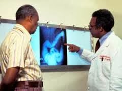
  • Imaging tests. An X-ray image of your lungs may reveal an abnormal mass or nodule. A CT scan can reveal small lesions in your lungs that might not be detected on an X-ray.
  • Sputum cytology. If you have a cough and are producing sputum, looking at the sputum under the microscope can sometimes reveal the presence of lung cancer cells.
  • Tissue sample (biopsy). A sample of abnormal cells may be removed in a procedure called a biopsy. Your doctor can perform a biopsy in a number of ways, including bronchoscopy, in which your doctor examines abnormal areas of your lungs using a lighted tube that's passed down your throat and into your lungs; mediastinoscopy, in which an incision is made at the base of your neck and surgical tools are inserted behind your breastbone to take tissue samples from lymph nodes; and needle biopsy, in which your doctor uses X-ray or CT images to guide a needle through your chest wall and into the lung tissue to collect suspicious cells. A biopsy sample may also be taken from lymph nodes or other areas where cancer has spread, such as your liver.
Once your lung cancer has been diagnosed, your doctor will work to determine the extent, or stage, of your cancer. Your cancer's stage helps you and your doctor decide what treatment is most appropriate.
Staging tests may include imaging procedures that allow your doctor to look for evidence that cancer has spread beyond your lungs. These tests include CT scans, magnetic resonance imaging (MRI), positron emission tomography (PET) and bone scans. Not every test is appropriate for every person, so talk with your doctor about which procedures are right for you.
Stages of lung cancer
  • Stage I. Cancer is limited to the lung and hasn't spread to the lymph nodes. The tumor is generally smaller than 2 inches (5 centimeters) across.
  • Stage II. The tumor at this stage may have grown larger than 2 inches, or it may be a smaller tumor that involves nearby structures, such as the chest wall, the diaphragm or the lining around the lungs (pleura). Cancer may also have spread to the nearby lymph nodes.
  • Stage III. The tumor at this stage may have grown very large and invaded other organs near the lungs. Or this stage may indicate a smaller tumor accompanied by cancer cells in lymph nodes farther away from the lungs.
  • Stage IV. Cancer has spread beyond the affected lung to the other lung or to distant areas of the body.
Small cell lung cancer is sometimes described as being limited or extensive. Limited indicates cancer is limited to one lung. Extensive indicates cancer has spread beyond the one lung.


Treatment for Lung Cancer at World Best Hospitals in India


Treatment of Lung cancer is depends on the type and stage of cancer. Lung cancer can be treated with surgery, chemotherapy, radiation therapy, or a combination of these treatments. The decision about which treatments will be appropriate for a given individual must take into account the location and extent of the tumor as well as the overall health status of the patient.

Surgery for Lung Cancer: If your cancer has not spread beyond the lungs, your general health is reasonably good and your breathing capacity is sufficient, the treatment that gives the best chance of cure is surgery. The most common operation is called a lobectomy, removes the affected part of the lung. Sometimes, the whole lung needs to be removed and this is called a pneumonectomy.
In patients with reduced lung function, smaller parts of the lung are removed to try to preserve breathing capacity. While these operations preserve breathing capacity, there is more likely to be a recurrence. Your doctor will advise you of the best operation for you.

Chemotherapy: This is the treatment of cancer with anti-cancer drugs. The aim is to destroy cancer cells while doing the least possible damage to normal cells. The drugs work by stopping cancer cells from multiplying. Chemotherapy is the treatment of choice for patients with small cell carcinoma. As this type of cancer spreads quickly, chemotherapy works well in treating it.

Radiotherapy: Radiotherapy treats cancer by using radiation to destroy or inhibit cancer cells. The radiation can be targeted onto cancer sites in your body. Treatment is carefully planned to do as little harm as possible to your normal body tissue.Treatment is painless and may take only two to three minutes. Usually, radiotherapy is given as a number of treatments over a period of weeks.

For more information visit:          http://www.medworldindia.com        
                    
https://www.facebook.com/medworld.india

Please scan and email your medical reports  to us at care@medworldindia.com and we shall get you a Free Medical Opinion from India’s Best Doctors.

Call Us : +91-9811058159
Mail Us : care@medworldindia.com


Friday 27 December 2013

Latest Radiotherapy for Prostate Cancer – CyberKnife Radiosurgery


The challenge that doctors face in treating prostate tumors with radiation therapy is that the prostate moves unpredictably as air passes through the rectum and as the bladder empties and fills. Minimizing any large movements of the prostate can help reduce unnecessary irradiation of surrounding healthy tissue. A new hope for patients who have inoperable or surgically complex tumors, or who may be looking for a non-surgical option.Cyberknife radiosurgery is a non –invasive, high-energy radiation treatment that delivers very precise beams of radiation from many angles outside the body. The accuracy of Cyberknife is so precise that radiation can be 'matched' to the shape of small complex tumors, even those located near critical organs. This ability allows Cyberknife to treat many lesions including some considered inoperable or untreatable with surgery.

 The CyberKnife Robotic Radiosurgery System is able to overcome this challenge by continuously identifying the exact location of the prostate and making active corrections for any movement of the prostate throughout the course of the treatment. During treatment, a patient lays still and breathes normally while the CyberKnife zeroes in on a moving target – the prostate – and irradiates it without harming surrounding areas. As a result, the procedure is more comfortable for patients, radiation is delivered more accurately and treatments can be completed in four to five days.

Currently the CyberKnifeRadiosurgery System is most frequently used by itself for patients with early stage prostate cancer confined to the prostate or in combination with another therapy, such as external beam radiation for patients with disease that extends beyond the prostate. Depending on the stage of the patient’s prostate cancer the doctor will recommend a treatment plan.  The CyberKnife System uses the fiducials as reference points to identify the exact location of the prostate. Doctors will wait approximately one week after insertion of the fiducials before CyberKnife treatment planning can begin to ensure that fiducial movement has stabilized.

 Prior to the treatment, a special custom-fit body cradle will be made. The cradle is made of a soft material that molds to the patient’s body, ensuring that the patient is in the same position for each treatment session and is comfortable during the procedure. While lying in the cradle, patients will undergo a CT scan. This CT data will be used by the CyberKnife team to determine the exact size, shape and location of the prostate. An MRI scan also may be necessary to fully visualize the prostate and nearby anatomy. Once the imaging is done, the body cradle will be stored and used during CyberKnife treatment.

For most patients, the CyberKnife treatment is a completely pain-free experience. They may dress comfortably in street clothes, and the CyberKnife center may allow patients to bring music to listen to during the treatment. Patients also may want to bring something to read or listen to during any waiting time, and have a friend or family member with them to provide support before and after treatment.


 When it is time for treatment, patients will lay on their custom body cradle. The radiation therapist will ensure the body cradle is properly adjusted and that patients are appropriately positioned on the treatment couch. When patients are ready for treatment to begin, the location of the prostate will be tracked and detected. The medical team will be watching patients every step of the way as the CyberKnife tracks the patient’s prostate as it moves, and safely and precisely delivering radiation to it. Once prostate cancer treatment is complete, most patients quickly return to their daily routines with little interruption to their normal activities. 


Saturday 21 December 2013

Nonsurgical Treatment for Liver Cancer: Extend Life and Improve Quality

Liver cancer or Hepatocellular carcinoma (HCC) is one of the commonest cancers in the world which have a high incidence of Hepatitis B infection. Apart from Hepatitis B, it may be caused by other diseases that lead to cirrhosis of the liver such as Hepatitis C infection, and alcohol abuse. 
Unfortunately, like many other cancers, liver cancer may go undetected until a late stage. It is often brought to attention by an ultrasound or CT scan done for pain in the upper abdomen or another unrelated symptom. Liver cancers are malignant tumors that grow on the surface or inside the liver. Typers of liver cancer are Hepatocellular Carcinoma , Cholangiocarcinoma  ,Hepatoblastoma .

Surgical removal of liver tumors offers the best chance for a cure. Unfortunately, liver tumors are often inoperable because the tumor may be too large, or has grown into major blood vessels or other vital structures. Sometimes, many small tumors are spread throughout the liver, making surgery too risky or impractical. Surgical removal is not possible for more than two-thirds of primary liver cancer patients and 90 percent of patients with secondary liver cancer.


There are a number of tests that can help in the diagnosis of cancer, including blood tests, physical examination and a variety of imaging techniques including X-rays (e.g., chest X-rays and mammograms); computed tomography (CT); magnetic resonance (MR) and ultrasound. Usually, however, the final diagnosis cannot be made until a biopsy is performed. In a biopsy, a sample of tissue from the tumor or other abnormality is obtained and examined by a pathologist. By examining the biopsy sample, pathologists and other experts also can determine what kind of cancer is present and whether it is likely to be fast or slow growing. This information is important in deciding the best type of treatment.
Although there are no standard or routine screening tests for liver cancer, the following tests are being used or studied to screen for it
  • Physical examination : Physical examination may show an enlarged, tender liver. Also, your skin and eyes may be checked for signs of jaundice.
  • Blood tests :Physical examination may show an enlarged, tender liver. Also, your skin and eyes may be checked for signs of jaundice.
  • Imaging Studies like Abdominal CT scan and Abdominal Ultrasound test
  • Liver biopsy or Aspiration.
  • Liver enzymes (liver function tests)
In order to check if the Liver Cancer has spread elsewhere in the body, the following tests are to be done:
  • PET Scan
  • Bone Scan- to check if the cancer has spread to the bones.


Tumors need a blood supply, which they actively generate, to feed themselves and grow. As vascular experts, interventional radiologists are uniquely skilled in using the vascular system to deliver targeted treatments via catheter throughout the body. In treating cancer patients, interventional radiologists can attack the cancer tumor from inside the body without medicating or affecting other parts of the body by using embolization and radiofrequency heat.

Embolization is a well-established interventional radiology technique that is used to treat trauma victims with massive bleeding, to control hemorrhage after childbirth, to decrease blood loss prior to surgery and to treat tumors. In treating cancer patients, interventional radiologists use embolization to cut off the blood supply to the tumor (embolization), deliver radiation to a tumor (radioembolization), or combine this technique with chemotherapy to deliver the cancer drug directly to the tumor (chemoembolization)

Chemoembolization is a minimally invasive treatment for liver cancer that can be used when there is too much tumor to treat with radiofrequency ablation (RFA), when the tumor is in a location that cannot be treated with RFA, or in combination with RFA or other treatments.
Chemoembolization delivers a high dose of cancer-killing drug (chemotherapy) directly to the organ while depriving the tumor of its blood supply by blocking, or embolizing, the arteries feeding the tumor. Using imaging for guidance, the interventional radiologist threads a tiny catheter up the femoral artery in the groin into the blood vessels supplying the liver tumor. The embolic agents keep the chemotherapy drug in the tumor by blocking the flow to other areas of the body. This allows for a higher dose of chemotherapy drug to be used, because less of the drug is able to circulate to the healthy cells in the body. Chemoembolization usually involves a hospital stay of two to four days. Patients typically have lower than normal energy levels for about a month afterwards.

Radiofrequency Ablation


For inoperable liver tumors, radiofrequency ablation (RFA) offers a nonsurgical, localized treatment that kills the tumor cells with heat, while sparing the healthy liver tissue. Thus, this treatment is much easier on the patient than systemic therapy. Radiofrequency energy can be given without affecting the patient's overall health and most people can resume their usual activities in a few days.
In this procedure, the interventional radiologist guides a small needle through the skin into the tumor. From the tip of the needle, radiofrequency energy (similar to microwaves) is transmitted to the tip of the needle, where it produces heat in the tissues. The dead tumor tissue shrinks and slowly forms a scar. The FDA has approved RFA for the treatment of liver tumors.


Targeted therapy is a treatment that targets the cancer’s specific genes, proteins, or the tissue environment that contributes to cancer growth and survival. This type of treatment blocks the growth and spread of cancer cells while limiting damage to normal cells. Recent studies show that not all tumors have the same targets. To find the most effective treatment, your doctor may run tests to identify the genes, proteins, and other factors in your tumor.

Benefits of Nonsurgical Treatment for Liver Cancer

  • Is most effective when all the cancer is localized in the liver
  • Can be used to treat primary liver cancer and tumors that have metastasized (spread) from other areas in the body to the liver
  • Usually does not require general anesthesia
  • Is well tolerated-most patients can resume their normal routine the next day and may feel tired for a few days
  • Can be repeated if necessary
  • May be combined with other treatment options
  • Can relieve pain and suffering for many cancer patients

For more information visit:          http://www.medworldindia.com       
                                                          
https://www.facebook.com/medworld.india

Please scan and email your medical reports  to us at care@medworldindia.com and we shall get you a Free Medical Opinion from India’s Best Doctors.

Call Us : +91-9811058159
Mail Us : care@medworldindia.com


The Most Successful Treatment for Rectal Cancer - Advanced Rectal Cancer Treatment

Rectal cancer occurs when cancerous cells develop in the tissue of the rectum. The rectum is the last part of the large intestine and leads to the anus, which is the opening to the outside of the body. Body waste is stored in the rectum until it is eliminated from the body through the anus.
Although rectal cancer is a life-threatening disease, it is a highly curable form of cancer if found early. Therefore, regular check-ups and screenings are very important.
Although the exact cause of rectal cancer is unknown, certain risk factors have been identified that may increase a person's chance of developing the disease.
·         A change in bowel habits
·         Diarrhea, constipation, or feeling that the bowel does not empty completely
·         Blood, either bright red or very dark in the stool
·         Stools that are narrower than usual
·         General abdominal discomfort such as frequent gas pains, bloating, fullness or cramps
·         Weight loss with no known reason

In making a diagnosis of rectal cancer, your doctor will first start by recording your medical history, asking about any symptoms you may be experiencing and conducting a thorough physical examination. He or she also may recommend one or more of the following diagnostic tests:
·         Digital Rectal Exam — This exam involves the doctor or nurse inserting a gloved, lubricated finger into the rectum to feel for an abnormalities.
·         Barium Enema — Also known as a lower gastrointestinal series, this test involves taking X-rays of the large intestines.
·         Fecal Occult Blood Test — This is a noninvasive test that detects the presence of hidden, or occult blood in the stool. Such blood may arise from anywhere along the digestive tract. Hidden blood in the stool is often the first, and in many cases the only, warning sign that a person has colorectal cancer.

Treatment for rectal cancer may include surgery, radiation therapy or chemotherapy, or a combination of these approaches.

Surgery

Surgery is the main treatment for all stages of rectal cancer, although radiation, chemotherapy, or both are often recommended in combination.
Some patients who undergo surgery for rectal cancer require a permanent colostomy — a surgically created opening in the abdominal wall through which waste is excreted. If you have a colostomy, our specially trained nurses will help you learn how to manage the colostomy and incorporate it into your lifestyle.
Depending on the location, stage and size of your tumor, your doctor will remove your cancer with one of the following methods:
·         Local excision — This surgical approach is used for very early stage cancers. It involves inserting a tube through the rectum into the colon and removing the cancer, rather than making a cut in the abdominal wall. If the cancer is found in a polyp, the procedure is called a polypectomy.
·         Resection and anastomosis — This approach is used for larger and more advanced cancers and involves removing the portion if the rectum containing the cancer, as well as the fatty tissue that surrounds the rectum and contains the lymph nodes. Afterwards, the doctor will sew the colon to the remaining rectum or the anus, during a procedure called an anastomosis.
·         Resection and colostomy — This approach is used when the rectum cannot be sewn back together. In these cases, a colostomy is performed, in which an opening outside of the body for waste to pass through is created, called a stoma. A bag is then placed around the stoma to collect the waste. The colostomy may be temporary, although if the entire rectum is removed, it is permanent. Our specially trained nurses will help you learn how to manage your colostomy and incorporate it into your lifestyle.

 Certain substances and drugs are used by the targeted therapy in order to target particular cancer cells without causing any harm to nearby normal cells. The two types of targeted therapy include :
·     
            Angiogenesis Inhibitors :This helps in stopping the growth of new blood vessels which tumors need to grow.
·      
        Monoclonal Antibodies : These antibodies are prepared in the laboratory with the help of a single type of immune system. Certain substances can be identified by these antibodies that are on the cancer cells or it also indentifies certain normal substances that are helping in the growth of cancer cells. These antibodies then attach themselves with the substances for destroying cancer cells and also prevent them from spreading.


ChemotherapyCertain types of drugs are used for stopping the growth of cancer cells or for destroying the cancer cells. The injection of these drugs is done into a vein from where they enter the bloodstream and they then travel through the entire body.

Radiation therapyHigh-energy beams are used for destroying cancer cells. Internal radiation and external radiation are the two types of radiation therapy. A radioactive substance that is sealed in catheters, needles wires or seeds is placed close to the cancer in the case of internal radiation. On the other hand, linear accelerators outside the body are used for sending radiation to the cancer in the case of external radiation. The type of radiation depends on the stage and type of the cancer.


·         World class results for Cancer Treatment
·         World Class equipment for investigations, radiotherapy and surgery
·         Cancer specialists with great qualifications and experience
·         India has many super specialists ( specialization in one particular area: Breast Cancers, Stomach Cancers, Prostate Cancers, etc)
·         Low cost of cancer treatment
·         India offers the perfect combination of expertise and economical costs


Friday 20 December 2013

Fight to End Cancer – World class results for Cancer Treatment in India

  • Stop smoking (active and passive) and alcohol to prevent lung, head and neck cancers.
  • Avoid high fat and rich foods; eat whole grains, fruits and fresh vegetables to prevent cancer of breast, prostate and colon.
  • Exercise to stay healthy
  • Avoid exposure to – household solvent cleaners, cleaning fluids, and paint thinners, pesticides, fungicides and other chemicals to avoid lung cancer.
  • Avoid multiple sex partners to avoid cancer cervix.
  • Avoid direct sunlight in summers to avoid skin cancers.
  • Get screening tests like the Pap smears, mammograms, Prostate Specific Antigen (PSA) and Digital Rectal Examinations (Dress) for secondary prevention.
  • Each and every individual should do self examination once a month.
  • Each and every individual visiting his / her doctor for any ailment should be screened for cancer.
  • Annual cancer check up should be done at cancer hospitals.
  • Suspected cancer cases should be investigated urgently to confirm or rule out cancer.
Steps before Starting the Treatment
  • Tissue diagnosis to confirm / rule out cancer by FNAC / Biopsy.
  • Staging work up to confirm stage of the disease.
  • Tumour Board Evaluation to chart out custom made treatment plan for each individual, as per standard treatment guidelines.
  • Choosing NABH Accredited cancer hospitals, offering quality cancer treatment.
  • A panel of cancer experts i.e. Medical Oncologists, Radiation Oncologists, Surgical Oncologists, Gynae Oncologists, Paediatric Oncologists, Onco-pathologists, Radiologists and Imaging experts, get together to review and plan suitable modality or modalities of treatment.
  • Treatment is need driven and not doctor driven.
  • Patients get the choice of surgery or radiotherapy, if the results are same by both modalities.
  • Patients are saved from unnecessary surgeries, if the surgeries cannot be performed.
  • Patients get complete treatment. This prevents recurrence due to incomplete treatment.
  • Chances of cure / improvement in quality of life are much higher because of right and optimum treatment.
  • Patients are saved from Medical shopping and get expert opinion under one roof by spending one consultation fee only.


Radiation
Cancer is treated by giving external radiation with linear accelerators and internal radiation i.e. brachytherapy. The newer techniques of Intensity Modulated Radiation Therapy ensures that healthy tissues surrounding the tumour are not exposed to radiation to prevent side effects of radiotherapy. Radiation oncologists and physicists work together as a team to give radiation.
Cancer Surgeries are very complex lasting 2 – 12 hours. A dedicated team of surgical oncologists and Anaesthesiologists, supported by other allied specialists is a must. Surgical oncologists are best suited to decide whether surgeries have to be conservative or radical.
Chemotherapies
Medical oncologists are trained exclusively to plan and manage chemotherapies and their side effects. Chemotherapies can be Normal and High Dose, including dose–intense and dose–dense; Infusional multiple and single drug; Bolus Chemotherapies; Intra-arterial chemotherapy; Targeted Therapies; Immunotherapy / Biological Therapies; Hormonal Therapies; Neutropenic Care; Nutritional Therapy; Palliative and Terminal Supportive Care; blood transfusion and I/V fluids etc. The radiotherapists and surgeons are not trained to perform chemotherapies.

Medworld India offers comprehensive care for patients with Cancer, including advanced diagnosis, best treatment options . A team of Surgical Oncologists, Radiation Oncologists, Medical Oncologists, Urologists, Rehabilitation team and other medical specialties work together to treat each Cancer patient We consider each patient's type and extent of Cancer to recommend the most appropriate treatment plan. They also carefully consider and select the treatment option that will allow the patient to maintain quality of life with good survival rate.

Why should you choose to get Indian hospitals offer the Best Cancer Treatment in India at affordable prices. MedWorld india associated Best Cancer Treatment Hospitals in India have the latest technology and infrastructure to offer the Most Advanced Cancer Treatment at low cost.

At MedWorld India Affiliated Best Cancer Hospitals are to deliver highest quality and advanced oncology care in a supportive and compassionate environment to all our patients, and to advance the treatment and prevention of cancers through innovative research.

MedWorld India Affiliated Best Cancer Hospitals in India offer:

  1.  World class results for Cancer Treatment
  2.  World Class equipment for investigations, radiotherapy and surgery
  3.  Cancer specialists with great qualifications and experience
  4.  India has many super specialists (specialization in one particular area: Breast      Cancers, Stomach Cancers, Prostate Cancers, etc)
  5.  Low cost of cancer treatment
  6.  India offers the perfect combination of expertise and economical costs

  For more information visit:          http://www.medworldindia.com      
                                                          
https://www.facebook.com/medworld.india

 Please scan and email your medical reports  to us at care@medworldindia.com and we shall get you a Free Medical Opinion from India’s Best Doctors.

  Call Us : +91-9811058159
  Mail Us : care@medworldindia.com


Contact Form

Name

Email *

Message *