Friday, 26 September 2014

Low Cost Breast Cancer Treatment and Surgery in India : Surgery for Breast Cancer

Planning Treatment for Breast Cancer - Surgery for Breast Cancer

Surgery usually involves removing part, or sometimes all, of the breast (mastectomy). The type of operation you have usually depends on the size and position of the cancer. Your surgeon will recommend surgery that keeps as much of the breast tissue and the shape of the breast as possible. This is called breast-conserving surgery. However, in some situations removing all of the breast (mastectomy) is advised. Your doctor and breast care nurse will talk you through your options.
Your surgery may also involve having the lymph nodes in your armpit removed or you may have tests to check the nodes. Sometimes women are given treatment with hormonal therapy or chemotherapy to shrink the cancer before they have surgery.
In early breast cancer, studies show that removing the lump followed by radiotherapy is as effective as a mastectomy. Some women may be asked to choose for themselves which operation to have

·         Wide local excision (lumpectomy)
·         Mastectomy

Wide local excision (lumpectomy)

This is when the cancer and an area of surrounding tissue is removed. It’s called a wide local excision or lumpectomy. This operation removes the affected breast tissue and for most women the appearance of their breast after a lumpectomy is good.
If the lump is very small, a fine wire (guide wire) is used to mark the area to be removed so that the surgeon can find it more easily. The doctor or radiologist will inject some local anaesthetic into the area to numb it before inserting the wire, using x-ray or ultrasound to guide them. 
Occasionally, an operation called a quadrantectomy is done. This removes a larger area of breast tissue. The effect on the appearance of the breast will be more noticeable than after a lumpectomy. The treated breast will be smaller than your other breast and there may be a noticeable dent in it. Women can have surgery to reduce the size of the other breast (mammoplasty) so that both breasts are the same size. 
After these operations you’ll need to have radiotherapy to the remaining breast tissue to destroy any cancer cells that may have been left behind.  

Some women may need to have all of the breast removed (mastectomy). A mastectomy is usually advised if:
·         the lump is large in proportion to the rest of the breast
·         there are areas of cancer in different parts of the breast (multi-focal)
·         there’s widespread DCIS in the breast
·         you’ve previously had radiotherapy to the chest, for example, to treat Hodgkin lymphoma.
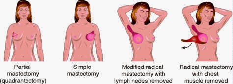
Mastectomy

Types of Mastectomy

A mastectomy removes all of the breast tissue. At the same time, you may have a test to check the lymph nodes in your armpit, or have some (or all) of the nodes removed.
A radicalmastectomy is where all the breast tissue, the muscles behind the breast and the lymph nodes in the armpit are removed. It’s only done if the cancer is found in the muscle under the breast. But this type of mastectomy is rarely needed as chemotherapy or hormonal therapy can usually be given before surgery to shrink the cancer. This means a mastectomy that removes only the breast tissue can then be done. 


Breastreconstruction

If you’re having a mastectomy, you’ll usually be offered breast reconstruction at the same time. This is when a new breast shape is formed. Breast reconstruction is very specialised surgery. Surgeons who do this type of operation may be plastic surgeons or oncoplastic surgeons, who are trained in both breast cancer surgery and reconstruction surgery.

Different techniques are used – for example, muscle from the back or the tummy area, or a silicone implant can be used. Some women may decide not to have it done immediately – it can be done months or even years after a mastectomy.

Medworld India offers comprehensive care for patients with Breast Cancer, including advanced diagnosis, best treatment options . A team of Surgical Oncologists, Radiation Oncologists, Medical Oncologists, Urologists, Rehabilitation team and other medical specialties work together to treat each Breast Cancer patient We consider each patient's type and extent of Breast Cancer to recommend the most appropriate treatment plan. They also carefully consider and select the treatment option that will allow the patient to maintain quality of life with good survival rate.

Why should you choose to get Indian hospitals offer the Best Cancer Treatment in India at affordable prices. MedWorld india associated Best Cancer Treatment Hospitals in India have the latest technology and infrastructure to offer the Most Advanced Cancer Treatment at low cost.;

At MedWorld India Affiliated Best Cancer Hospitals are to deliver highest quality and advanced oncology care in a supportive and compassionate environment to all our patients, and to advance the treatment and prevention of cancers through innovative research.

MedWorld India Affiliated Best Cancer Hospitals in India offer:

·         World class results for Cancer Treatment
·         World Class equipment for investigations, radiotherapy and surgery
·         Cancer specialists with great qualifications and experience
·         India has many super specialists ( specialization in one particular area: Breast Cancers, Stomach Cancers, Prostate Cancers, etc)
·         Low cost of cancer treatment
·         India offers the perfect combination of expertise and economical costs
 
 For more information visit:          http://www.medworldindia.com     
                                                          
https://www.facebook.com/medworld.india
Please scan and email your medical reports  to us at care@medworldindia.com and we shall get you a Free Medical Opinion from India’s Best Doctors.
·          
·         Call Us : +91-9811058159
·         Mail Us : care@medworldindia.com
·          

Wednesday, 24 September 2014

Advanced Technology for Brain Tumor : Best Cancer Hospitals in India offer the World's Advanced Brain Tumor Treatment in India

Indian Hospital are providing comprehensive medical and surgical care for patients with disorders of the brain, brain tumor surgery is routinely carried out with results at par with the best centers globally. As we all know that India is now becoming a medical hub and a growing destination for Brain Tumor surgery. Hospitals of brain tumor surgery India provides international quality of medical healthcare services by the best brain surgeons in India who are equipped with the most advanced medical treatment and brain tumor healing techniques. 

Brain tumor is one an abnormal mass of tissue in which cells grows and multiply, seemingly unchecked by the mechanisms that control normal cells. Indian Neurologists and Neurosurgeons are supported by the most extensive neuro-diagnostic and imaging facilities including Asia's most advanced MRI and CT technology. Along with providing general diagnostic X-ray imaging, Hospitals of brain tumor surgery in India offers you a magnitude of imaging services like EEG, EMG, Sensation 10 CT Scanner, Functional MRI with Spectro-scopy, OPMI Multivision etc.

Treatment

People diagnosed with a CNS tumor generally need to seek treatment as soon as possible. The pressure caused by a growing CNS tumor can cause severe symptoms, including a backup of CSF and problems with blood circulation, which can damage delicate nerves and deprive cells of nourishment.

Treating brain and spinal cord tumors can be difficult. The blood-brain barrier, which normally serves to protect the brain and spinal cord from damaging chemicals entering those structures, also keeps out many types of potentially beneficial chemotherapeutic drugs. Surgery can be difficult if the tumor is near a delicate portion of the brain or spinal cord. Radiation therapy can damage healthy tissue. However, research in the past two decades has improved the survival rates of patients with brain tumors. More refined surgeries, a better understanding of what types of tumors respond to chemotherapy, and precise delivery of radiation have resulted in a longer life span and better quality of life for patients with brain tumors.

Surgery

Surgery is the most common type of treatment for a brain tumor and is often the only treatment performed for a benign brain tumor. Even if the cancer cannot be cured, its removal can relieve symptoms if it is creating pressure on parts of the brain. There have been rapid advances in surgery for a brain tumor, including the use of cortical mapping and enhanced imaging devices to give surgeons more tools to plan and perform the surgery. For a tumor that is near the speech center, it is increasingly common to perform the operation under awake conditions. Typically, the patient is awakened once the surface of the brain is exposed, and special electrical stimulation techniques are used to locate the speech center and thereby avoid causing any damage while removing the tumor.

Surgery to the brain requires the removal of part of the skull, a procedure called a craniotomy. After the surgeon removes the tumor, the patient's own bone will be used to cover the opening in the skull. Gliadel wafers that deliver chemotherapeutic drugs (see below) require surgery to put the wafers in the tumor bed site. This may be done at the same time as a craniotomy.

In addition to removing or reducing the size of the brain tumor, surgery can provide a tissue sample for analysis. For some tumor types, the results of the analysis can help in showing if chemotherapy or radiation therapy will be useful.

Radiation therapy

Radiation therapy is the use of high-energy x-rays or other particles to kill cancer cells. Oncologists may use radiation therapy along with surgery to slow the growth of aggressive tumors. Radiation can be directed in the following ways:

The linear-accelerator machine provides external-beam radiation therapy from outside the body to target the tumor within the brain; this is becoming increasingly more precise with the addition of multileaf collimators (a device that restricts and confines the x-ray beam to a given treatment area).

In stereotactic radiosurgery, a computer assembles images from CT scans or MRI scans to locate the tumor and help direct the radiation. This may involve the use of an external head device to "marry" the patient's head/tumor location to the incoming radiosurgery beams.

Brachytherapy uses radioactive seeds implanted directly in the tumor site; however, this treatment technique is rarely used at this time for a brain tumor.

Radiation techniques

The following radiation techniques may be used:

Conventional radiation therapy. The treatment location is determined based on anatomic landmarks and x-rays. In certain situations, such as whole brain radiation therapy for brain metastases, this technique is appropriate. For more precise targeting, more elaborate techniques are required. Chemotherapy may be used in conjunction with this type of radiation therapy.

Three-dimensional conformal radiation therapy. Based on CT and MRI images, a three-dimensional model of the tumor and normal tissues is created on a computer. Beam size and angles are determined that maximize tumor dose and minimize normal tissue dose.

Stereotactic radiosurgery. Stereotactic radiosurgery involves delivering a single, high dose of radiation directly to the tumor and not healthy tissues. It works best for a tumor that is only in one area of the brain and certain benign tumors, but is also used for multiple metastatic brain tumors. There are three methods by which stereotactic radiosurgery is performed:
  • A modified linear accelerator is a machine that creates high-energy radiation by using electricity to form a stream of fast-moving subatomic particles.
  • A gamma knife is another form of radiation therapy that concentrates highly focused beams of gamma radiation on the tumor.
  • A cyber knife is a robotic device used in radiation therapy to guide radiation to the tumor target-particularly targets in the brain, head, and neck regions.
Fractionated stereotactic radiation therapy. Radiation therapy is delivered with stereotactic precision but divided into small daily fractions over several weeks using a relocatable head frame, in contrast to the one-day radiosurgery. This technique is used for tumors located close to eloquent or sensitive structures, such as the optic nerves or brain stem.

Intensity modulated radiation therapy (IMRT). Radiation therapy is delivered with greater intensity or dose to thicker areas of the tumor and with less intensity to thinner areas of the tumor. This is accomplished by placing tiny metal leaves in the beam to reduce the intensity of the beam in order to customize the shape of the dose to the shape of the tumor.

All of these more elaborate techniques are designed to achieve greater precision and minimize the dose to the surrounding normal brain tissue. Depending on the size and location of the tumor, the radiation oncologist may choose any of the above radiation techniques. In certain situations, a combination of two or more techniques is appropriate.
Very young children (younger than 5) are not usually appropriate candidates for radiation therapy because of high risk of damage to their developing brains.

Chemotherapy

Chemotherapy is the use of drugs to kill cancer cells. The goal of chemotherapy can be to destroy cancer remaining after surgery, slow the tumor's growth, or reduce symptoms.

Advanced/recurrent brain tumors

If, in spite of initial treatment, the brain tumor does not go into remission (the temporary or permanent disappearance of symptoms) or if it recurs, patients can still receive care to manage the symptoms caused by the tumor. Symptom management is always important since the symptoms of a brain tumor can interfere with quality of life.

Since a brain tumor is so rare, it can be hard for doctors to plan treatments unless they know what has worked in treating other patients with a brain tumor. Clinical trials are research studies that compare the standard treatments (the best treatments available) with newer treatments that may be more effective. Investigating new treatments involves careful monitoring using scientific methods and all participants are followed closely to track progress.

Due to advances in research, new drugs are being created to combat brain tumors. Many of these new drugs are called "small molecules" or "molecularly targeted therapies" because they are small in size (and can therefore be taken orally) and/or can attack a specific molecule or target within the brain tumor cells. These new drugs are being tested either alone or in combination with standard chemotherapy


Medical tourism in India is a boon for abroad patients who are searching for cost effective brain tumor surgery in India operated by the best Brain surgeons. Medical tourism companies has been providing valuable information and guidance regarding neurosurgery or brain tumor surgery in India to international patients as the number of patients having brain disorders have started coming to the neuro surgeons of India for brain tumor surgery at an affordable price. When it comes to price, abroad patients consider India as the best place for this surgery. Medical tourism has broad appeal as it is providing best medical healthcare facilities during this brain tumor surgery in India at very low cost as compare to western countries. 

International patients are looking forward to India just because of first class medical facilities at third class rates. Many of India's surgery hospitals and clinics offer international-standard brain tumor surgery in India at local prices, no waiting list and a high quality holiday. India is a land of art and culture and it is always seemed that abroad people are always attracted to it because of its variety.  
For more details on brain tumor surgery in India visit : www.medworldindia.com


Friday, 19 September 2014

Endometrial Cancer: Screening & Prevention; Symptoms; Treatment Options in India


The most common cancer of a reproductive system of a woman is known as uterine cancer. This cancer starts in cell layers that form the lining of the uterus. This cancer can be detected in its early stage as it causes abnormal vaginal bleeding. The surgical removal of the uterus can cure uterine cancer.


Types of Uterine Cancer :- 

Sarcoma: Sarcoma cancer grows in the supporting tissues of the uterine glands or in the myometrium. This cancer accounts for 2-4% of uterine cancers.

Adenocarcinoma: This cancer is considered as the main type of uterine cancer that grows from the cells in the uterus lining. Endometrial cancer is the other name of this cancer.

  • Unusual Vaginal Bleeding or Discharge
  • Trouble Urinating
  • Pelvic Pain
  • Pain during sexual intercourse
  • Discomfort or pain in the lower abdomen
Diagnosis of Endometrial Cancer (Uterine Cancer)


A pelvic exam is performed by the doctors checking the vagina, uterus, ovaries, bladder, and rectum for Endometrial Cancer (Uterine Cancer). An instrument called a speculum is used to widen the vagina so the doctor can see the upper portion of the vagina and the cervix.

The Pap Smear test is often performed during a pelvic exam Endometrial Cancer (Uterine Cancer). The doctor uses a wooden scraper (spatula) or small brush to collect a sample of cells from the cervix and upper vagina. The cells are then sent to a medical laboratory to be checked for abnormal changes. Because uterine cancer begins inside the uterus, it may not show up on a Pap test, which examines cells from the cervix.

A biopsy may be needed for a definitive diagnosis where the doctor removes a sample of tissue from the uterine lining. In some cases, a woman may require a dilation and curettage (D&C), which is usually a day care procedure. During a D&C, the opening of the cervix is widened and the doctor scrapes tissue from the lining of the uterus. A pathologist examines the tissue to check for cancer cells, hyperplasia, or other conditions.

Common Endometrial Cancer (Uterine Cancer) Symptoms
  •   Abnormal bleeding- heavy bleeding between periods, or heavy bleeding  during irregular periods
  •    Pain during intercourse, pelvic pain or pain in the legs or back
  •    Difficulty urinating or pain during urination
  •    Enlarged uterus , Vaginal discharge and or unexplained weight loss
The following are the stages of endometrial cancer

Stage I of endometrial cancer

In  stage I, cancer is found in the uterus only. Stage I is divided into stages IA, IB, and IC, based on how far the cancer has spread.
  • Stage IA: Cancer is in the endometrium only.
  • Stage IB: Cancer has spread into the inner half of the myometrium.
  • Stage IC: Cancer has spread into the outer half of the myometrium.
Stage II of endometrial cancer 
In stage II, cancer has spread from the uterus to the cervix, but has not spread outside the uterus. Stage II is divided into stages IIA and IIB, based on how far the cancer has spread into the cervix.
  • Stage IIA: Cancer has spread to the glands where the cervix and uterus meet.
  • Stage IIB: Cancer has spread into the connective tissue of the cervix.
Stage III of endometrial cancer

In stage III, cancer has spread beyond the uterus and cervix, but has not spread beyond the pelvis. Stage III is divided into stages IIIA, IIIB, and IIIC, based on how far the cancer has spread within the pelvis.
  • Stage IIIA: Cancer has spread to one or more of the following:
  • The outermost layer of the uterus; or
  • Tissue just beyond the uterus; or
  • The peritoneum.
  • Stage IIIB: Cancer has spread beyond the uterus and cervix, into the vagina.
  • Stage IIIC: Cancer has spread to lymph nodes near the uterus.
Stage IV of endometrial cancer

In stage IV, cancer has spread beyond the pelvis. Stage IV is divided into stages IVA and IVB, based on how far the cancer has spread.
  • Stage IVA: Cancer has spread to the bladder and/or bowel wall.
  • Stage IVB: Cancer has spread to other parts of the body beyond the pelvis, including lymph nodes in the abdomen and/or groin.

Surgical Treatment of EndometrialCancer (Uterine Cancer)

Surgery is the best option when Endometrial Cancer (Uterine Cancer) is diagnosed in its very early stages. At this time, the location and the stage of cancer make removal easy. Sometimes patients are treated with a combination of surgery and radiation therapy. Laparoscopy Assisted Vaginal Hysterectomy - involves the minimally invasive or open surgery to remove the uterus. If required teh surgeon may decide to remove both ovaries and the fallopian tubes if there is a risk of Endometrial cancer spread to the ovaries.

In most cases, uterine cancer is an Adenocarcinoma that metastasizes late, usually from the endometrium to the cervix, ovaries, fallopian tubes, and other peritoneal structures. It may spread to distant organs, such as the lungs and the brain, through the blood or the lymphatic system. In such cases advanced tratement post Hystrectomy like Chemotherapy and Radiation are advised

Chemotherapy 

Involves modern cancer drugs that are administered intravenously or orally to kill cancer cells and to reduce the chances of the tumour returning elsewhere in the body.


Radiation therapy 

India's leading Cancer hospitals now have some of the world's moset advanced radiation equipment like Linac, Novelis, Gamma Knife and Cyberknife for treating Colon rectal Cancers. High technology radiation is used to kill cancer cells without affecting the healthy tissues. Radiation may be used to reduce the tumor prior to surgery or to obviate the symptoms of colorectal cancer such as pain, bleeding, or blockage.

For more information visit:          http://www.medworldindia.com      
                    
https://www.facebook.com/medworld.india

Please scan and email your medical reports  to us at care@medworldindia.com and we shall get you a Free Medical Opinion from India’s Best Doctors.

Call Us : +91-9811058159


Mail Us : care@medworldindia.com

Tuesday, 9 September 2014

Prostatectomy Surgery Options in India : Types of Prostatectomy

A surgical approach to treating prostate cancer will remove all or part of the prostate. Typically, men with early-stage disease or cancer that’s confined to the prostate will undergo radical prostatectomy—removal of the entire prostate gland, plus some surrounding tissue. Other surgical procedures may be performed on men with advanced or recurrent disease.

In the most common type of prostatectomy—radical retropubic prostatectomy—an incision is made in the abdomen and the prostate is removed from behind the pubic bone. The surgeon then stitches the urethra directly to the bladder so urine is able to flow. 
Because it typically takes a few days for the body to get used to this new setup, the surgeon will insert a catheter, or tube, into the bladder. Urine will flow automatically out of the bladder, down the urethra, and into a collection bag without the need for conscious control of the sphincter. The catheter is usually kept in place for about a week to 10 days.
Another type of surgery, known as radical perineal prostatectomywhich includes removal of tissues near the gland, usually cures prostate cancer if the surgery removes all the malignant cells. The incision is usually made through the lower abdomen (retropubic) but also can be made in the groin (perineal). Typically, general anesthesia is used and the patient remains in the hospital for two to four days following surgery. Full recovery may require one month or more. 

Partial prostatectomy
 is generally a treatment for enlargement of the prostate, which is common after age 50. It can also be used to reduce pain and ease urination in men with advanced prostate cancer.

Complications from prostatectomy are those associated with any surgery, including infection and bleeding. Other complications vary depending on the age and health of the man, type of surgery and experience of the surgeon. Two primary concerns men have about radical prostatectomy are the possibilities of impotence and incontinence. Patients can retain sexual function if nerves are not injured during the surgery. Urinary incontinence is usually temporary but bladder problems can continue for years. Radical prostatectomy does cause sterility, but there are procedures that may allow a man to father children, if desired.

Procedure

Before the procedure the bladder and urethra may be numbed (local anesthesia), body below the waist may be numbed (regional anesthesia), or the patient may be completely asleep (general anesthesia). The procedure itself usually takes about 45 minutes. The doctor inserts a cystoscope (a thin, tubelike telescope) through the urethral opening in the penis and into the urethra. The doctor can view the urethra and prostate, either through the cystoscope or on a video monitor. The laser is inserted through the cystoscope to the area of the prostate. The laser is then used to destroy excess prostate tissue.

Following prostatectomy will vary depending on the type of surgery performed: -

  • Radical prostatectomy (retropubic or perineal) : - This surgery may require the patient to stay hospitalized for two or three days and miss work for three to five weeks. The catheter may remain for one to three weeks. When the patient urinates, the fluid flows through the plastic tube and collects in a bag for disposal.
  • Laparoscopic prostatectomy : - Long-term experience with laparoscopic prostatectomy is not yet available. This surgery may require a hospital stay of only one day. Some physicians and patients report that it involves fewer complications, less pain and faster recovery than open surgery.

  • Partial (simple) prostatectomy (transurethral resection of the prostate [TURP] or transurethral incision of the prostate [TUIP]) : - Generally, a patient is discharged from the hospital after a day or two and can return to work in a week or two. A catheter placed through the urethra is used for a few days to drain urine from the bladder. TUIP does not always require a hospital stay.
For more information visit:          http://www.medworldindia.com       
                    
https://www.facebook.com/medworld.india

Please scan and email your medical reports  to us at care@medworldindia.com and we shall get you a Free Medical Opinion from India’s Best Doctors.

Call Us : +91-9811058159
Mail Us : care@medworldindia.com

Friday, 5 September 2014

Most Advanced Laparoscopic Surgery to Treat Colon Cancer

Colon cancer is cancer of the large intestine (colon), the lower part of your digestive system. Rectal cancer is cancer of the last several inches of the colon. Together, they're often referred to as colorectal cancers. Most cases of colon cancer begin as small, noncancerous (benign) clumps of cells called adenomatous polyps. Over time some of these polyps become colon cancers.

These are the third most commonly diagnosed cancers in world. They start as a small proliferated, ulcerated lesions or thickenings from the inner most layers of bowel lining. Left untreated they grow in size, extend through the wall to adjacent organs, involve nearby lymph nodes and at latter stages involve the liver and other distant organs, through blood stream, manifesting as different stages of disease progression.


Many cases of colon cancer have no symptoms. The following symptoms, however, may indicate colon cancer:
·         Pain and tenderness in lower abdomen
·         Passing of Blood in the stool
·         Constipation, Diarrhea or other changes in bowel habits like narrow stools.
·         Abrupt Weight loss

How is for Colon Cancer diagnosed?

·         Colonoscopy: An endoscope is inserted into the rectum and advanced through the colon, through this the doctor can examine the entire colon.
·         Diagnosis is confirmed with a colon biopsy - Stage of disease is confirmed by pathologists and imaging tests, such as computerized tomography (CT or CAT) scans.
·         Endoscopic ultrasound and magnetic resonance imaging (MRI) may also be used to stage rectal cancer
·         Sigmoidoscopy: In this an endoscope is interested in the rectum and moved through the left side of the colon. It cannot be used to view the middle and right sides of the colon.
·         Fecal occult blood test (FOBT) along with Complete blood count (CBC to check for anemia and CT , MRI or PET scans of the abdomen, pelvic area.

Stages of Colon Cancer

The staging of colon cancer helps doctors determine a course of treatment. Staging is done on a scale from 0 to 4, with the higher stages indicating a cancer than has spread more throughout the body.

·         Stage 0: In this stage, the cancer hasn’t grown beyond the inner layer of the colon or rectum.
·         Stage I: The cancer has spread through the colon’s inner lining, but hasn’t spread beyond the colon wall or rectum.
·         Stage II: The cancer has grown through the colon or rectal wall, but hasn’t spread to nearby lymph nodes.
·         Stage III: The cancer has invaded nearby lymph nodes but isn’t affecting other parts of the body.
·         Stage IV: The cancer has metastasized throughout the body to sites such as the liver, lung, ovaries or the lining of the abdominal cavity.


Surgery to treat colon cancer

Surgery is the cornerstone of treatment for colon cancer. You may need to have the entire colon removed or only part of your colon.
·     How much of your colon has to be removed depends on the location and particular characteristics of your tumour. Sometimes only a polyp is cancerous, and removal of the polyp may be all that is necessary.
·      
      Surgery may also be done to relieve symptoms when the cancer has caused a bowel obstruction. The usual procedure is bypass for obstructions that cannot be cured.
Rather than making long incisions common in traditional "open" surgery, laparoscopic surgery requires only small incisions to perform the operation.

What is laparoscopic surgery?

The word "laparoscopy" means to look inside the abdominal cavity with a special camera or scope. To perform laparoscopy, three or four small (five-10mm) incisions are made in the abdomen. The laparoscope and surgical instruments are inserted through these small incisions. The surgeon is then guided by the laparoscope, which transmits a picture of the intestinal organs on a video monitor.
The benefit of using this surgical approach is that you are less likely to experience pain and scarring after surgery, a more rapid recovery and less risk of infection.


Colon surgeons have used laparoscopy to treat the following conditions:
·         Crohn's disease
·         Certain bowel cancers
·         Diverticulitis
·         Familial polyposis
·         Faecal incontinence
·         Rectal prolapse


·        

Tuesday, 2 September 2014

A study suggests having chemotherapy before surgery to remove a tumor may benefit more esophageal cancer patients

A study suggests having chemotherapy before surgery to remove a tumor may benefit more patients with esophageal cancer than previously thought.
Previous studies have already established that giving chemotherapy before surgery can benefit patients with esophageal cancer by shrinking their tumors.
But as well as these patients, the new study shows that even those whose tumors do not shrink may also benefit, because the pre-op chemo can reduce the spread of the cancer to the lymph nodes. The study shows that when spreading to the lymph nodes was reduced, this had a knock-on effect of improving survival and lengthening time to relapse.

We still don't know why some patients respond to chemotherapy, and others don't. But this study suggests that more patients than previously thought do benefit from chemotherapy before surgery, which means that having more treatment after their operation might also work for them too.He and his colleagues also believe having more chemo after surgery would benefit such patients even further.

An aggressive cancer with a very poor prognosis

Esophageal cancer is an aggressive cancer with a very poor prognosis. The cancer forms in the lining of the esophagus or gullet, the pipe that passes food from the throat to the stomach.The cancer is more common in people who smoke, drink alcohol, are overweight and whose diet is poor in fresh fruits and vegetables.

The  Cancer Institute estimates that 18,980 people will be diagnosed with the disease and 16,210 will have died of it in 2014

In the UK, where around 8,500 new cases are diagnosed every year, esophageal cancer is the ninth most common cancer, and the sixth most common cause of cancer death.

Study investigated 200 patients with adenocarcinoma

There are two types of esophageal cancer, squamous cell carcinoma, which starts in the flat cells in the gullet lining, and adenocarcinoma, which starts in cells that make and release fluids like mucus.

Average time to relapse extended from 1 to over 5 years


However, in the patients who had the pre-op chemo but whose tumors did not respond, there were 30 individuals who showed a reduction in the number of lymph nodes with cancer in them. And this group showed an increase in the average time to relapse from just under 1 year to over 5.5 years.
                                Most Advanced Esophageal Cancer Care and Treatment in India 

Medworld India 
offers comprehensive care for patients with Esophageal Cancer, including advanced diagnosis, best treatment options . A team of Surgical Oncologists, Radiation Oncologists, Medical Oncologists, Urologists, Rehabilitation team and other medical specialties work together to treat each Esophageal Cancer patient We consider each patient's type and extent of Esophageal Cancer to recommend the most appropriate treatment plan. They also carefully consider and select the treatment option that will allow the patient to maintain quality of life with good survival rate.


Why should you choose to get Indian hospitals offer the Best Cancer Treatment in India at affordable prices. MedWorld india associated Best Cancer Treatment Hospitals in India have the latest technology and infrastructure to offer the Most Advanced Cancer Treatment at low cost.

At MedWorld India Affiliated Best Cancer Hospitals are to deliver highest quality and advanced oncology care in a supportive and compassionate environment to all our patients, and to advance the treatment and prevention of cancers through innovative research.

MedWorld India Affiliated Best Cancer Hospitals in India offer:
  • World class results for Cancer Treatment
  • World Class equipment for investigations, radiotherapy and surgery
  • Cancer specialists with great qualifications and experience
  • India has many super specialists ( specialization in one particular area: Breast Cancers, Stomach Cancers, Prostate Cancers, etc)
  • Low cost of cancer treatment.
  • India offers the perfect combination of expertise and economical costs

Contact Form

Name

Email *

Message *